
Pre-Clinical Micro-CT
Pre-Clinical Micro-CT
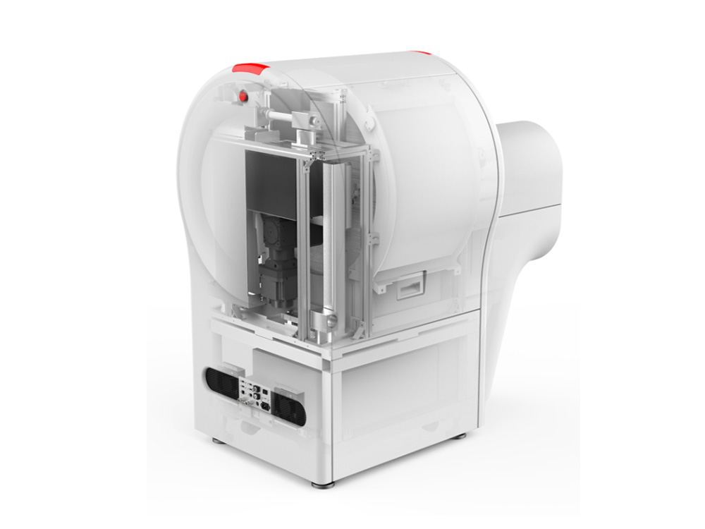
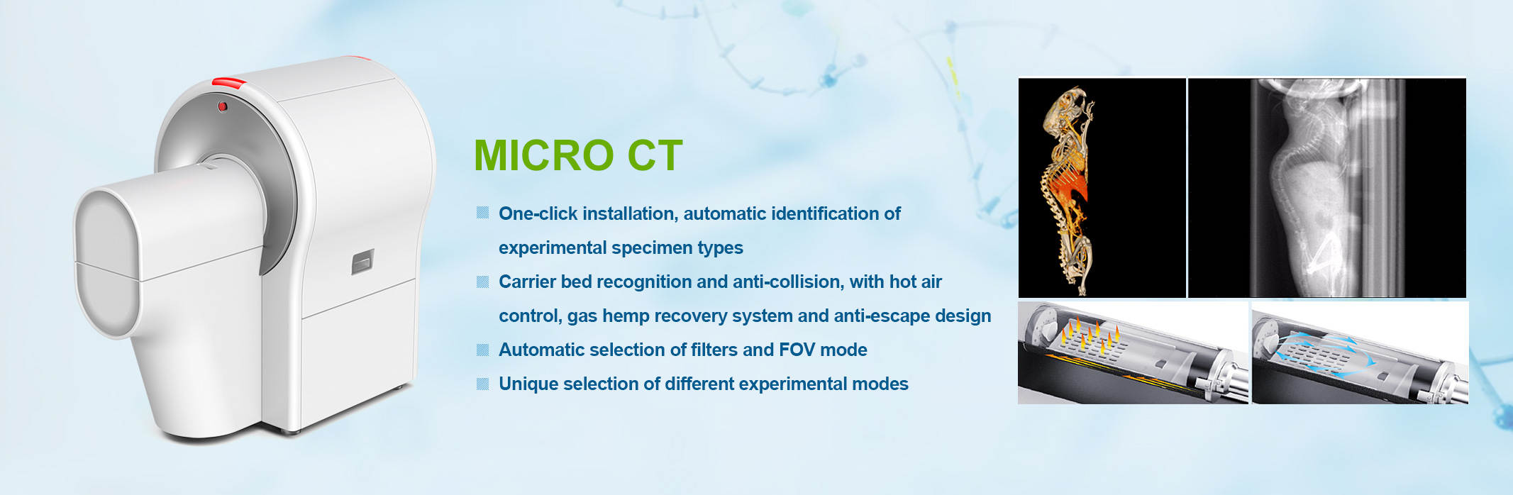
Model: DELab μCT-100
DELab μCT-100 is the first microcomputer tomography scanner made in Taiwan. Except for X-ray tubes and imagers, all components and software development are designed and manufactured by the Delta team. The main application areas include new drugs and disease development In the field of preclinical animal experiments, agriculture, industry.
Product description:
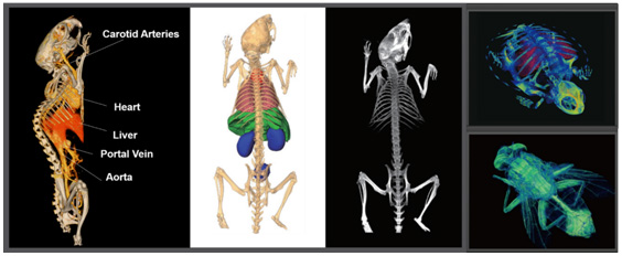
This Micro-CT can scan the whole body of the experimental animal (including bone, fat, tumor, blood vessels, whole body organs, etc.). At the same time, the software supporting the system is easy to operate and has a high quantitative function, which can provide quantitative information of dozens of parameters such as bone measurement, fat rate, distance, area, and volume.
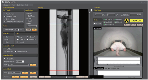
1. Multi-mode operating system (high-resolution mode, mouse and ex vivo experiment mode and rat experiment mode), automatic detection of test specimens, and automatic configuration of test parameters.
2. Intelligent scanning mode: the experiment is preset at one time, without the need for professional personnel or long-term use training.
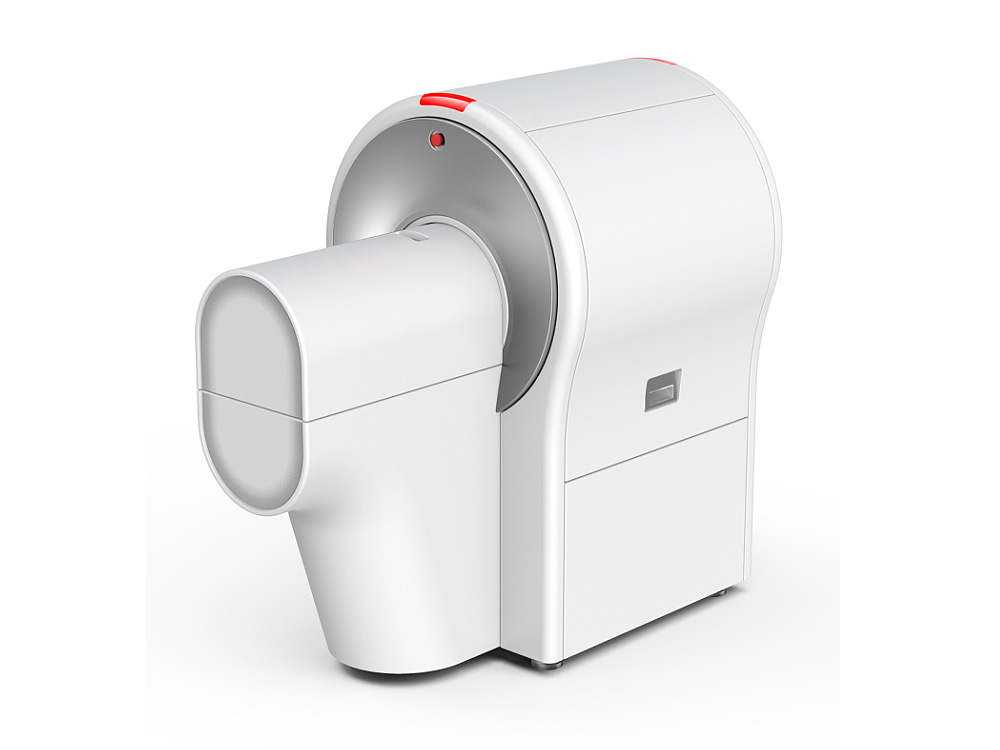
3. One-click loading bed installation, the system automatically recognizes the loading bed and quickly sets the filter and FOV mode selection.
4. The fastest scan time for a test is 2 seconds (high-speed mode), and the animal is exposed to little radiation.
5. Continuous scanning can be performed to shorten the scanning time, protect animals, and increase the utilization rate of the instrument.
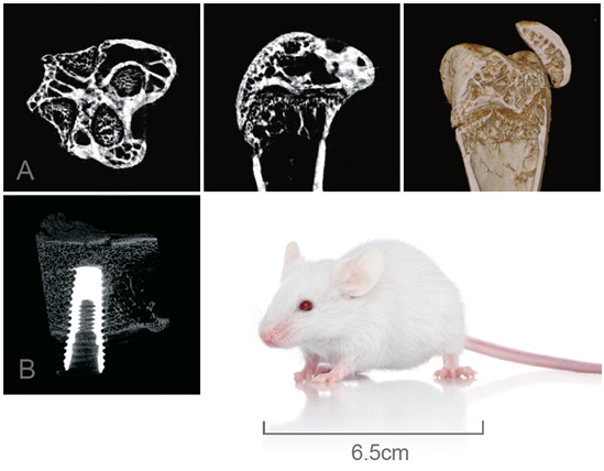
6. The highest 3D resolution of the system can reach 2μm, high-resolution imaging.
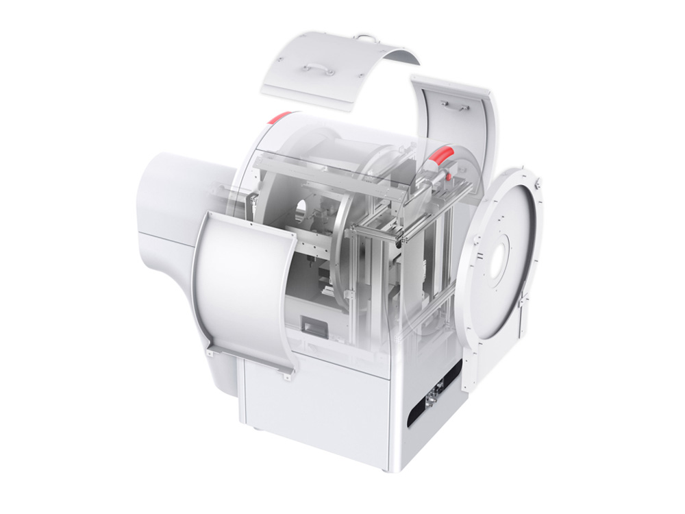
7. Different experimental specimens are equipped with different carrier beds. The carrier beds are equipped with animal escape shields, which are safer to use.
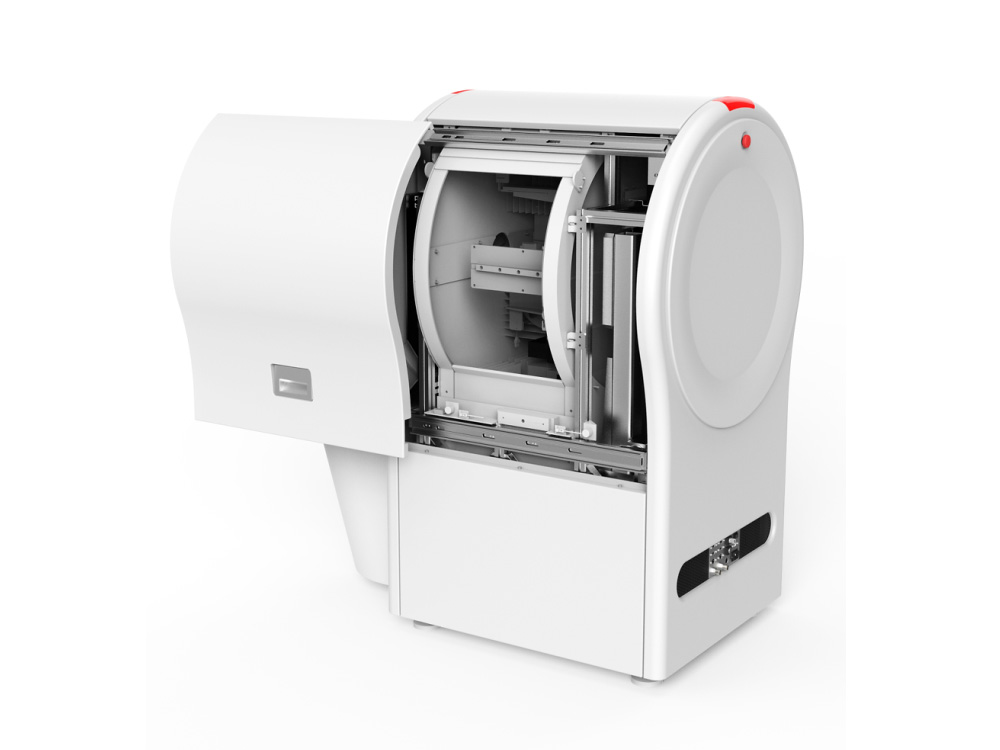
8. Multifunctional carrier bed system, equipped with gas anesthesia, ECG, body temperature, camera, adjustable hot air system, participating in gas anesthesia recovery system.
9. The bed cover has anti-pinch design, so it is more reliable to use.
10. The CT is equipped with 6 metal filters, and the filters are automatically configured, which is safe to use and reduces damage to laboratory animals.
11. Experimental conditions can be customized and saved.
12. High-precision platform, hardware mechanical calibration, get more accurate and clear experimental results.
13. The instrument comes with a lead shielding room, and the maximum dose is 10 cm away from the surface of the instrument. The radiation dose is less than 1 μSv / h
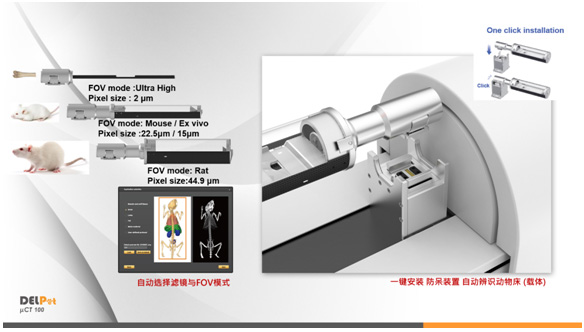
ONE touch automatically recognizes the carrier bed and quickly sets the selection filter and FOV mode:
Suitable forthe bone / mouse / rat scan:
The industry's fastest scanning speed 2 seconds: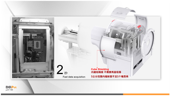
Preclinical small animal research:
Study on isolated femur and titanium alloy:
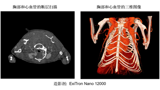
Tumor vascular research:
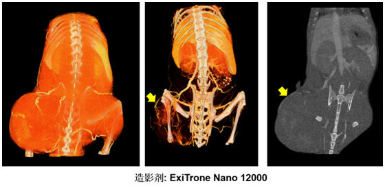
Cardiovascular Research:
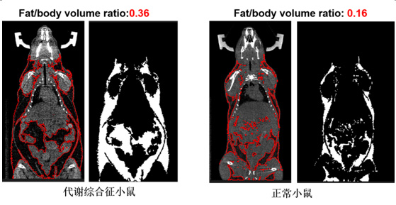
Fat Metabolism Research / Analysis:
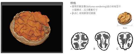
Food Research:
Technical Parameters:
1. X-ray source (maximum energy): 40-90kV, 50W
2. Extra filter: the system randomly gives 6 filter
3. X-ray imager: 1536 x 1944, 14-bit CMOS detector, 2x2, 4x4 merge mode
4. Pixel size: best up to 2 μm
5. Effective range of CT imaging: 80 x 200 mm
6. Scan time: standard scan 20 seconds, fast mode 2 seconds
7. The acquisition software and analysis software developed by the company can scan and analyze bones, fats, tumors, blood vessels, and organs of the whole body. Quantitative information such as bone density, fat rate, distance, area, and volume can be measured.
8. Software post-processing:
Data interface: DICOM, JPEG, TIFF, BMP, RAW and other formats;
Two-dimensional processing of image file input and output: image browsing, selection, processing and display, two-dimensional geometric transformation and measurement, continuous image playback, etc .;
Three-dimensional processing: three-dimensional tissue segmentation, surface model reconstruction and layered display, slice reorganization;
Three-dimensional display: photorealistic display, lighting parameter adjustment, three-dimensional geometric transformation, three-dimensional measurement;
Image segmentation: provide multiple segmentation methods (Thresholding, Otsu ...)
9. Power supply: AC 100-220V / 10A / 50-60Hz
10. Operating temperature: 10-30 ℃
11. Operating humidity: <85%, no condensation
12. The instrument reserves USB port, HDMI port, can connect to the Internet
13. The instrument is equipped with X-ray emergency stop switch
14. The instrument is equipped with a door machine safety interlock, and anti-trapping and other safety devices
15. The computer and host equipment are integrated, and do not occupy additional space
16. Give a personal radiation dose detector at random.
CONTACT US
CONTACT US
Add:Room 416, incubation base, 280 Waihuan East Road, Xiaoguwei street, Panyu District, Guangzhou
Contact:Manager Luo Phone:15360620228 Tel:020-39921409 Email:sail@lsolg.com | 
Follow Us |